A Conserved Developmental Patterning Network Produces Quantitatively Different Output in Multiple Species of Drosophila
Charless Fowlkes, Kelly Eckenrode, Meghan Bragdon, Miriah Meyer, Zeba Wunderlich, Lisa Simirenko, Cris Luengo, Soile Keränen, Clara, Henriquez, David Knowles, Mark Biggin, Michael Eisen, Angela DePace
Differences in the level, timing, or location of gene expression can contribute
to alternative phenotypes at the molecular and organismal level. Understanding
the origins of expression differences is complicated by the fact that
organismal morphology and gene regulatory networks could potentially vary even
between closely related species. To assess the scope of such changes, we used
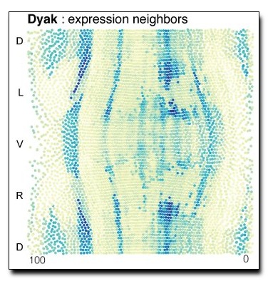
high-resolution imaging methods to measure mRNA expression in blastoderm
embryos of Drosophila yakuba and Drosophila pseudoobscura and assembled these
data into cellular resolution atlases, where expression levels for 13 genes in
the segmentation network are averaged into species-specific, cellular
resolution morphological frameworks. We demonstrate that the blastoderm embryos
of these species differ in their morphology in terms of size, shape, and number
of nuclei. We present an approach to compare cellular gene expression patterns
between species, while accounting for varying embryo morphology, and apply it
to our data and an equivalent dataset for Drosophila melanogaster. Our analysis
reveals that all individual genes differ quantitatively in their
spatio-temporal expression patterns between these species, primarily in terms
of their relative position and dynamics. Despite many small quantitative
differences, cellular gene expression profiles for the whole set of genes
examined are largely similar. This suggests that cell types at this stage of
development are conserved, though they can differ in their relative position by
up to 3–4 cell widths and in their relative proportion between species by as
much as 5-fold. Quantitative differences in the dynamics and relative level of
a subset of genes between corresponding cell types may reflect altered
regulatory functions between species. Our results emphasize that
transcriptional networks can diverge over short evolutionary timescales and
that even small changes can lead to distinct output in terms of the placement
and number of equivalent cells.
Download: pdf
Text Reference
Charless Fowlkes, Kelly Eckenrode, Meghan Bragdon, Miriah Meyer, Zeba Wunderlich, Lisa Simirenko, Cris Luengo Hendriks, Soile Keränen, Clara, Henriquez, David Knowles, Mark Biggin, Michael Eisen, and Angela DePace.
A conserved developmental patterning network produces quantitatively different output in multiple species of drosophila.
PLoS Genetics, 7:e1002346, 2011.
BibTeX Reference
@article{Fowlkes_PLOS_2011,
AUTHOR = {Fowlkes, Charless and Eckenrode, Kelly and Bragdon, Meghan and Meyer, Miriah and Wunderlich, Zeba and Simirenko, Lisa and Luengo Hendriks, Cris and Ker{\"a}nen, Soile and Henriquez, Clara, and Knowles, David and Biggin, Mark and Eisen, Michael and DePace, Angela},
TITLE = "A Conserved Developmental Patterning Network Produces Quantitatively Different Output in Multiple Species of Drosophila",
JOURNAL = "PLoS Genetics",
YEAR = "2011",
VOLUME = "7",
ISSUE = "10",
PAGES = "e1002346",
TAG = "biological_images"
}